
NAD+ vs NADH: Oxidized and Reduced Forms Compared for Research
NAD+ and NADH are the oxidized and reduced forms of the same coenzyme, nicotinamide adenine dinucleotide. They differ at one site: the nicotinamide ring. In NAD+ the ring is an aromatic pyridinium that can accept a hydride; in NADH it carries that hydride at carbon 4, as a 1,4-dihydronicotinamide. Dehydrogenases transfer the hydride at C-4 stereospecifically, and NAD(P)-linked enzymes make up about 17% of classified enzymes (You, 1985). The NAD+/NADH couple has a standard reduction potential (E0') of about -320 mV (Buckel and Thauer, 2018).
That one-ring difference changes how the two forms absorb light, fluoresce, survive acid and buffer, and behave during extraction. This page compares them for assay design and handling. Only NAD+ is in the catalog; NADH is discussed as its reduced partner and is not sold here. For NAD+ against its salvage-pathway precursors, see NAD+ vs NMN.
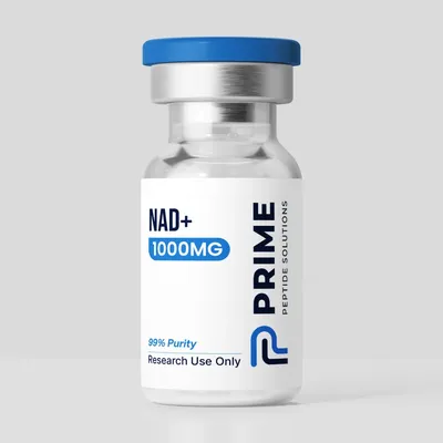
The research-grade NAD+ supplied by Prime Peptide Solutions is sold strictly for in-vitro laboratory research. It is not for human or animal consumption.
| Property | NAD+ | NADH |
|---|---|---|
| Redox state | Oxidized; hydride acceptor | Reduced; hydride donor |
| Nicotinamide ring | Aromatic pyridinium | 1,4-dihydropyridine (hydride at C-4) |
| PubChem record | CID 5892 (nadide) | CID 439153; disodium salt CID 12598256 |
| Formula and MW | C21H27N7O14P2; 663.4 g/mol | C21H29N7O14P2; 665.4 g/mol (disodium salt C21H27N7Na2O14P2, 709.4 g/mol) |
| Monoisotopic mass | 663.109 Da | 665.125 Da |
| CAS | 53-84-9 | 58-68-4 |
| UV absorbance | 260 nm; molar absorptivity 17.4 × 10³ L mol-1 cm-1 | 260 and 340 nm; 14.1 × 10³ at 260 nm and 6,317 at 340 nm |
| Fluorescence | Readouts use the reduced form | Fluorescent; lifetime about 0.4 ns free and 1.0 ns enzyme-bound |
| Acid | Recovered in acid extracts | Acid-catalyzed hydration and degradation |
| Buffer stability (43 days) | Highly stable in Tris | Most stable in Tris; faster loss in phosphate and HEPES |
| Free ratio in rat liver | About 725 NAD+ per NADH in cytoplasm; about 8 in mitochondria | |
| In this catalog | 1000mg vial | Not sold |
About NAD+ (the oxidized form)
Identity. PubChem files NAD+ as nadide, CID 5892: C21H27N7O14P2, 663.4 g/mol, monoisotopic mass 663.109 Da, CAS 53-84-9. It is the form that accepts a hydride from a substrate in dehydrogenase reactions.
Spectroscopy. Using purified NADH as the reference, one study set the molar absorptivity of NAD+ at 260 nm at 17.4 × 10³ L mol-1 cm-1 at 25 °C (Haid et al., 1975). Its reduction is followed by the rise in absorbance at 340 nm, which belongs to the NADH that forms (McComb et al., 1976).
In solution. In a 43-day study at 19 and 25 °C, NAD+ was highly stable in Tris buffer (Wolfe et al., 2024). Classic extraction methods recover the oxidized form from an acid extract and the reduced form from a separate basic extract (Klaidman et al., 1995); one comparison used 0.6 M perchloric acid for NAD+ and 0.5 M potassium hydroxide in 50% ethanol for NADH (Bessho et al., 1989).
About NADH (the reduced form)
Identity. PubChem lists NADH under CID 439153 (synonym 1,4-dihydronicotinamide adenine dinucleotide): C21H29N7O14P2, 665.4 g/mol, monoisotopic mass 665.125 Da, CAS 58-68-4. The disodium salt has its own record, CID 12598256 (C21H27N7Na2O14P2, 709.4 g/mol). The neutral formulas differ by two hydrogens, about 2.016 Da.
Absorbance at 340 nm. From 85 independent determinations, the molar absorptivity of NADH at 340 nm is 6,317 L mol-1 cm-1 at 25 °C and pH 7.8 (McComb et al., 1976). Purified NADH gave a 260/340 nm absorbance ratio of 2.265; using 6.22 × 10³ at 340 nm, that ratio gives a molar absorptivity at 260 nm of 14.1 × 10³ (Haid et al., 1975).
Fluorescence. Reduced pyridine nucleotide fluorescence was used as early as 1962 to record redox changes in rat kidney and brain in situ (Chance et al., 1962). Fluorescence lifetime imaging separates free from enzyme-bound NADH: decay times were about 0.4 ns free and 1.0 ns when bound to malate dehydrogenase (Lakowicz et al., 1992).
Acid and buffers. NADH undergoes acid-catalyzed hydration. Studied between pH 1 and 7, the rate depended on hydrogen-ion concentration at moderate pH and became independent of it at low pH; the reaction is general-acid catalyzed, most buffers raise the rate as their concentration rises, and beta-NADH also anomerizes to the alpha form (Johnson and Tuazon, 1977). A kinetic study of NADPH, with NADH included for comparison, found degradation rates that rose with temperature, hydronium ion, phosphate and acetate, and NADPH was generally less stable than NADH under the same conditions (Wu et al., 1986). At 340 nm, NADH degraded faster in phosphate than in PIPES buffer, which the authors assigned to a phosphate adduct with the pyridine ring, and they found mildly alkaline PIPES buffer at low temperature the better condition (Rover Junior et al., 1998). Over 43 days, NADH in Tris lost 4 µM per day at 19 °C and 11 µM per day at 25 °C (more than 90% and 75% left), while phosphate and HEPES reached up to 34 µM per day; the loss proceeded through oxidation and de-aromatization of the dihydropyridine ring and could be followed by UV-visible spectroscopy (Wolfe et al., 2024).
Which Form Fits the Research Question?
NAD+ and NADH are not interchangeable reagents. The choice follows the direction of the reaction and the readout:
- Dehydrogenase assays that reduce the coenzyme start from NAD+ and read the appearance of NADH at 340 nm, as in the coupled assay used to measure the NADH absorptivity (McComb et al., 1976). Assays run the other way start from NADH and read its loss.
- Enzymes that consume NAD+ need the oxidized form as substrate.
- Fluorescence and imaging work relies on NADH, the fluorescent form; lifetime methods separate its free and bound pools.
- Measuring the NAD+/NADH ratio in cells or tissue is mostly an extraction problem. Interconversion between the oxidized and reduced forms during extraction was a major barrier; an acidic 40:40:20 acetonitrile:methanol:water mix with 0.1 M formic acid gave the least interconversion, extracts had to be neutralized at once to avoid acid-catalyzed degradation, and isotope labeling plus spiked standards were used to track interconversion (Lu et al., 2018). A single-sample method lysed yeast under nitrogen in cold ammonium acetate and separated both forms by HPLC with UV detection (Sporty et al., 2008).
- Interpreting a ratio requires knowing which pool was measured. In rat liver, the free NAD+/NADH ratio was about 725 in the cytoplasm and about 8 in mitochondria, and totals differ from free ratios because much more NADH than NAD+ is protein-bound (Williamson et al., 1967).
- Long incubations or cell-free systems: in the 43-day buffer study, Tris gave the slowest NADH losses and phosphate or HEPES the fastest.
Related pages: NAD+ vs NMN, MOTS-C vs NAD+, NAD+ vs Glutathione, the NAD+ research overview and HPLC and mass spec explained.
Key Studies
- Williamson et al., 1967 (Biochem J): free NAD+/NADH ratios in cytoplasm and mitochondria. PubMed 4291787
- Haid et al., 1975 (Clin Chem): molar absorptivities of NADH and NAD at 260 nm. PubMed 165910
- McComb et al., 1976 (Clin Chem): molar absorptivity of NADH at 340 nm. PubMed 2388
- Johnson and Tuazon, 1977 (Biochemistry): acid-catalyzed hydration of NADH. PubMed 14674
- Lakowicz et al., 1992 (PNAS): fluorescence lifetimes of free and bound NADH. PubMed 1741380
- Lu et al., 2018 (Antioxid Redox Signal): extraction without interconversion. PubMed 28497978
- Wolfe et al., 2024 (Molecules): 43-day stability of NAD+ and NADH in common buffers. PubMed 39598842
Frequently Asked Questions
What is the difference between NAD+ and NADH?
NAD+ is the oxidized form and NADH the reduced form of nicotinamide adenine dinucleotide. NADH carries an extra hydride at carbon 4 of the nicotinamide ring, which NAD+ accepts from substrates in dehydrogenase reactions.
How can NAD+ and NADH be told apart in solution?
By absorbance at 340 nm, where NADH has a molar absorptivity of about 6,317 L mol-1 cm-1, by NADH fluorescence, or by mass: the neutral forms differ by about 2 Da. HPLC separates the two before UV detection.
Which form is more sensitive to acid and buffer?
NADH. It undergoes acid-catalyzed hydration, and its degradation rate rises with hydronium ion, phosphate and temperature. In a 43-day study both forms were highly stable in Tris, with faster NADH losses in phosphate and HEPES.
Why do published NAD+/NADH ratios vary so much?
Because extraction can convert one form into the other, free and protein-bound pools differ, and cytoplasm and mitochondria hold very different ratios. One method tracked interconversion with isotope labeling and spiked standards.
How should the NAD+ powder be handled?
Keep the vial tightly closed, cold, dry and away from light, and let it reach room temperature before opening. In solution, buffer and temperature govern stability, as summarized above. General conditions are in the storage guide.
Is NADH sold here, and what is the NAD+ intended for?
NADH is not sold here. The research-grade NAD+ is sold strictly for in-vitro laboratory research and is not for human or animal consumption. Published lab reports (COAs) are listed on our COAs page.
Shop NAD+
- Ships the same business day
- Free US shipping on $199.99+
- 14-day money-back guarantee on damaged items