

Why Receptor Signaling Matters for GLP-1 Research
Every GLP-1 receptor agonist, from native GLP-1 to long-acting analogs, acts through the same protein: the glucagon-like peptide-1 receptor (GLP-1R). Ligands differ not only in how tightly they bind but in which intracellular pathways they activate, for how long, and what happens to the receptor afterward. Those differences are measurable with standard cell-based assays.
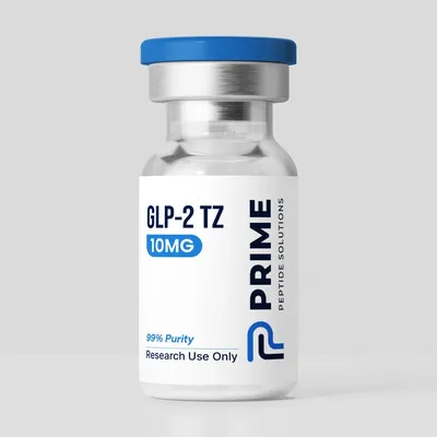
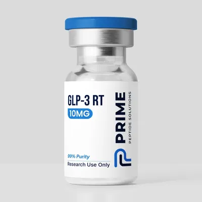
This article explains the receptor itself: how peptides bind it, the Gαs-cAMP pathway that couples it to insulin secretion in beta cells, the role of beta-arrestin in internalization and desensitization, and the concept of biased agonism. Compound-specific mechanisms are covered separately in our semaglutide, tirzepatide and retatrutide overviews, and the catalog naming of these compounds is explained in GLP-1, GLP-2 and GLP-3 naming explained.
GLP-1R: A Class B1 GPCR
GLP-1R is a class B G protein-coupled receptor (GPCR), the secretin-like family (often called class B1) that also includes the receptors for glucagon, GIP, GLP-2, secretin, PTH and calcitonin. Class B receptors share two structural features: a large N-terminal extracellular domain (ECD) and the seven-transmembrane (7TM) core common to all GPCRs (Underwood et al., 2010; Zhang et al., 2018).
The two-domain binding model
Peptide ligands for class B receptors are described by a two-domain binding model: the C-terminal part of the peptide is captured by the ECD, and the N-terminal part reaches down into a pocket formed by the transmembrane helices, where it triggers activation (Zhang et al., 2018). The ECD acts largely as an affinity trap, while the N-terminus does the signaling work.
The first half of this model was visualized in crystal structures of the isolated ECD. Underwood and colleagues solved the GLP-1R ECD bound to GLP-1 at 2.1 Å resolution and found that GLP-1 forms a kinked but continuous alpha-helix from Thr13 to Val33 when bound to the ECD. Their mutagenesis data also suggested that GLP-1 and exendin-4 bind the full-length receptor in somewhat different ways (Underwood et al., 2010). The second half required full-length receptor structures, which arrived with cryo-EM.
What cryo-EM showed
In 2017, Zhang and colleagues reported a near-atomic cryo-EM structure of GLP-1R bound to GLP-1 and the stimulatory G protein Gs. The peptide was clasped between the N-terminal domain and the transmembrane core and further stabilized by the extracellular loops. On the intracellular side, activation produced a sharp kink in the middle of transmembrane helix 6, swinging its intracellular half outward to make room for the α5-helix of Gαs (Zhang et al., 2017). A crystal structure of the related glucagon receptor bound to a peptide agonist added detail on the stalk region and first extracellular loop, which changed conformation on peptide binding, leading the authors to extend the two-domain model into a "dual-binding-site trigger" model (Zhang et al., 2018).
The Gαs-cAMP Pathway in Beta Cells
GLP-1R signals primarily through Gs (Zhang et al., 2017). Once the receptor adopts its active conformation, it catalyzes GDP-GTP exchange on Gαs, which then stimulates membrane adenylyl cyclase and raises intracellular cyclic AMP (cAMP). In pancreatic beta cells, cAMP acts through two main effectors:
- Protein kinase A (PKA): the classic cAMP-dependent kinase, which phosphorylates targets involved in ion channel activity and the exocytotic machinery.
- Epac2 (cAMP-GEFII): a cAMP-binding guanine nucleotide exchange factor that activates the small GTPase Rap1 and interacts with Rim2, acting independently of PKA.
Both arms contribute. In isolated islets, Kashima and colleagues reported that knocking down Epac2 with antisense oligonucleotides, or blocking PKA with H-89, each reduced incretin-potentiated insulin secretion by about 50%, while combining the two reduced it by roughly 80-90% (Kashima et al., 2001). Shibasaki and colleagues later showed in Epac2-knockout mice that cAMP-potentiated granule fusion during the first phase of glucose-induced exocytosis was markedly reduced, and that Rap1, activated through Epac2, mediated cAMP-induced insulin secretion in a PKA-independent manner (Shibasaki et al., 2007).
Why the effect is glucose-dependent
A defining property of GLP-1R signaling in beta cells is that it amplifies insulin secretion rather than triggering it. Glucose metabolism closes ATP-sensitive potassium channels, depolarizes the cell and opens voltage-gated calcium channels; cAMP then potentiates the calcium-triggered exocytosis. In the Shibasaki study, cAMP signaling alone did not cause granule fusion but potentiated both the first and second phases of glucose-induced fusion events (Shibasaki et al., 2007), and the Epac2-Rim2 effect described by Kashima depended on intracellular calcium as well as cAMP (Kashima et al., 2001). This is why GLP-1 action is described as glucose-dependent insulin secretion (Underwood et al., 2010; Drucker, 2018).
Beta-Arrestin, Internalization and Desensitization
Like most GPCRs, an activated GLP-1R is phosphorylated on its intracellular C-terminal tail, which promotes binding of beta-arrestin 1 or beta-arrestin 2. Beta-arrestin binding has three broad consequences in the GPCR literature:
- Desensitization: arrestin sterically competes with G protein coupling, damping further cAMP generation from receptors at the plasma membrane.
- Internalization: arrestin links the receptor to the clathrin endocytic machinery, removing it from the cell surface. Internalized receptors are then sorted toward recycling or degradation.
- Scaffolding: arrestin can organize signaling complexes of its own, for example involving ERK.
For GLP-1R specifically, the picture is nuanced. Sonoda and colleagues reported in INS-1 beta cells that beta-arrestin-1 co-immunoprecipitated with GLP-1R, and that knocking down beta-arrestin-1 reduced GLP-1-stimulated cAMP, ERK and CREB activation and insulin secretion. Notably, the knockdown did not change receptor surface expression or ligand-induced internalization in their system (Sonoda et al., 2008). Later work pointed the other way for sustained signaling: in primary islets, beta-arrestin-1 limited the insulin response to GLP-1 (Willard et al., 2020). Arrestin's role therefore appears to depend on cell type, timescale and readout, so researchers measure several pathways in parallel.
Biased Agonism at GLP-1R
Biased agonism (also called functional selectivity) describes ligands that stabilize receptor conformations favoring one downstream pathway over another, compared with a reference agonist. At GLP-1R the usual comparison is Gαs-cAMP signaling versus beta-arrestin recruitment, with native GLP-1 as the reference.
The structural basis
Wootten and colleagues used analytical methods that separate pathway-specific signaling from effects on affinity, combined with receptor mutagenesis, and concluded that peptide agonists can engage different elements of the GLP-1R extracellular face to achieve effector coupling and biased signaling (Wootten et al., 2016). Liang and colleagues then solved a 3.3 Å phase-plate cryo-EM structure of GLP-1R bound to the G protein-biased peptide exendin-P5 and Gαs. Compared with the GLP-1-bound structure, it differed in the arrangement of extracellular loop 3 and nearby transmembrane segments, and showed a six-degree difference in the angle at which the Gαs α5-helix engaged the receptor (Liang et al., 2018).
Exendin-phe1 and receptor trafficking
A widely cited example comes from Jones et al., 2018. The group studied a series of biased GLP-1R agonists derived from exendin-4 with different tendencies to drive receptor internalization and recycling; the best-known is exendin-phe1, which carries a phenylalanine substitution at position 1. Compared with a panel of approved GLP-1 mimetics, compounds that kept GLP-1R at the plasma membrane produced greater long-term insulin release in their assays. The authors linked this to reduced beta-arrestin recruitment and faster agonist dissociation rates, and reported glycemic effects in mice without a concomitant increase in signs of nausea (Jones et al., 2018).
Tirzepatide's cAMP-biased GLP-1R profile
Tirzepatide is a dual GIP and GLP-1 receptor agonist. In a pharmacology study from Willard et al., 2020, tirzepatide mimicked native GIP at the GIP receptor but showed bias at the GLP-1 receptor, favoring cAMP generation over beta-arrestin recruitment. This coincided with a weaker ability than GLP-1 to drive GLP-1R internalization. In primary islets, beta-arrestin-1 limited the insulin response to GLP-1 but not to GIP or tirzepatide, which the authors interpreted as the biased profile enhancing insulin secretion. The same paper described the molecule as "imbalanced," with greater engagement of the GIP receptor than the GLP-1 receptor in their occupancy analysis (Willard et al., 2020). Cryo-EM structures of tirzepatide bound to GIPR and GLP-1R have since mapped the interactions behind this dual activity (Zhao et al., 2022).
Related Receptors: GIPR and the Glucagon Receptor
GLP-1R has two close relatives that matter for multi-agonist research. Both are class B1 GPCRs that couple to Gαs and follow the two-domain binding model:
ReceptorEndogenous ligandPrimary G proteinExample agonists in researchGLP-1RGLP-1GαsSemaglutide, tirzepatide, retatrutideGIPRGIPGαsTirzepatide, retatrutideGCGRGlucagonGαsRetatrutide, peptide 20
Zhao and colleagues determined cryo-EM structures of tirzepatide bound to GIPR and GLP-1R, and of the triagonist peptide 20 bound to GIPR, GLP-1R and GCGR, revealing both shared and receptor-specific interactions behind dual and triple agonism (Zhao et al., 2022). The glucagon receptor crystal structure discussed above shows how the ECD and transmembrane domain reorient relative to each other during peptide binding (Zhang et al., 2018).
How Labs Measure GLP-1R Signaling
Bias, desensitization and trafficking are inferred from concentration-response curves in engineered cell lines (commonly HEK293 or CHO cells expressing the human receptor) and in beta-cell models such as INS-1 cells or isolated islets. The main readouts are:
cAMP accumulation assays
These quantify cAMP after agonist exposure, typically with a phosphodiesterase inhibitor present. Common formats include homogeneous time-resolved FRET (HTRF) immunoassays, luminescent biosensors and FRET or BRET cAMP sensors that report changes in real time. Readouts are potency (EC50) and maximal response (Emax) relative to native GLP-1.
Beta-arrestin recruitment (BRET and complementation)
Bioluminescence resonance energy transfer (BRET) assays fuse a luciferase to the receptor and a fluorescent acceptor to beta-arrestin, or the reverse; arrestin recruitment increases energy transfer. Split-luciferase complementation works on the same proximity principle. Running these alongside cAMP assays in the same cells allows a bias factor to be calculated.
Internalization and trafficking assays
Receptor endocytosis is tracked with epitope- or self-labeling tag approaches (for example SNAP-tagged receptors labeled with a cell-impermeant fluorophore), surface ELISA, high-content imaging, or BRET between the receptor and markers of the plasma membrane or endosomes. Recycling is measured by following the return of labeled receptors after agonist washout.
Downstream and functional readouts
ERK1/2 phosphorylation, CREB activation and, in beta-cell models, glucose-stimulated insulin secretion at basal and elevated glucose complete the picture. Measuring insulin secretion at low and high glucose is how glucose dependence is shown experimentally.
Interpreting bias carefully
Apparent bias depends on the reference ligand, the cell line, receptor expression level, assay kinetics and the amplification inherent in each readout; cAMP assays are highly amplified, arrestin recruitment assays much less so. Methods that separate affinity from efficacy, as in Wootten et al., 2016, and time-resolved measurements help avoid labeling a ligand as biased when it simply differs in potency or binding kinetics.
Frequently Asked Research Questions
Does cAMP alone trigger insulin release?
No. In the beta-cell studies cited here, cAMP potentiated glucose-induced exocytosis but did not cause granule fusion on its own, which underlies the glucose dependence of GLP-1R action (Shibasaki et al., 2007).
Is beta-arrestin always inhibitory at GLP-1R?
Not uniformly. One study in INS-1 cells found beta-arrestin-1 supported GLP-1 signaling to insulin secretion (Sonoda et al., 2008), while work in primary islets found it limited the insulin response to GLP-1 (Willard et al., 2020). Context and timescale matter.
Conclusion
GLP-1R is a class B1 GPCR that binds peptides through its extracellular domain and transmembrane core, signals mainly through Gαs and cAMP to PKA and Epac2, and amplifies insulin secretion only in the presence of glucose. Beta-arrestin recruitment and internalization shape how long that signal lasts, and ligands such as exendin-phe1 and tirzepatide show that these pathways can be pulled apart. Cryo-EM structures of GLP-1R, GIPR and GCGR now give a structural framework for that pharmacology, and paired cAMP, arrestin and trafficking assays are how laboratories measure it.
Disclaimer: This article is provided for educational and research purposes only. It summarizes publicly available scientific literature and does not constitute medical advice. Semaglutide, tirzepatide, retatrutide and all peptide compounds sold by Prime Peptide Solutions are intended strictly for laboratory research, are not approved for human use, and are not for human or animal consumption. Researchers are responsible for compliance with all applicable regulations in their jurisdiction.
Shop these products
- Ships the same business day
- Free US shipping on $149.99+
- 7-day money-back guarantee on damaged items
References & Further Reading
- PubMed: GLP-1 receptor biased agonism
- PubMed: GLP-1 receptor, beta-arrestin and internalization
- PubMed: Epac2 and cAMP-potentiated insulin secretion
- PubMed: Class B GPCR and GLP-1R cryo-EM structures
- Comparison: Semaglutide vs tirzepatide vs retatrutide
Research-Grade GLP-1 Receptor Agonists
In our catalog, semaglutide is listed as GLP-1 SM, tirzepatide as GLP-2 TZ (10mg and 20mg vials) and retatrutide as GLP-3 RT, each supplied as a lyophilized powder with bacteriostatic water sold separately. Published batch Certificates of Analysis are on our COA page.
Sold strictly for in-vitro laboratory research. Not for human or animal consumption.