

What Is Thymosin Alpha-1?
Thymosin alpha-1 is a 28-amino-acid peptide with an acetylated N-terminus, studied in immunology research for its links to toll-like receptor (TLR) signaling in dendritic cells and to T-cell maturation pathways. It is the N-terminal segment of a larger nuclear protein, prothymosin alpha, and its sequence was first determined in 1977 from a calf-thymus extract known as thymosin fraction 5.
Despite the shared name, thymosin alpha-1 is unrelated to thymosin beta-4 and is not TB-500; thymosin alpha-1 and thymosin beta-4 come from different genes and protein families. This overview covers its structure, origin, reported signaling pathways and laboratory handling.
This article is a scientific overview for laboratory researchers. The research-grade thymosin alpha-1 supplied by Prime Peptide Solutions is sold strictly for in-vitro laboratory research. It is not approved for human use and is not for human or animal consumption.
Structure and Background
Sequenced from thymosin fraction 5
A 1977 paper in PNAS determined the amino-acid sequence of a polypeptide isolated from calf thymus and named it thymosin alpha-1. The authors described a highly acidic molecule of 28 residues, one of several peptides in thymosin fraction 5 that may participate in the regulation, differentiation and function of thymus-dependent lymphocytes (T cells). The same paper suggested a nomenclature for the family of polypeptides present in fraction 5.
Prothymosin alpha, the precursor
A 1984 PNAS study isolated a larger polypeptide from rat thymus, prothymosin alpha, which carries the thymosin alpha-1 sequence at its N-terminus. It concluded that prothymosin alpha appears to be the native polypeptide from which thymosin alpha-1 and other fragments are generated during the isolation of thymosin fraction 5.
UniProt (entry P06454, gene PTMA) annotates thymosin alpha-1 as residues 2-29 of human prothymosin alpha, a nuclear protein of the pro/parathymosin family. The initiator methionine is removed and serine 2 carries an N-acetyl group, which becomes the acetylated N-terminus of the peptide. A 2023 review describes cleavage by asparagine endopeptidase, and a 2007 review states that the peptide is produced in vivo by cleavage of prothymosin alpha in diverse mammalian tissues, not in the thymus alone.
Molecule at a Glance
- Sequence: Ac-SDAAVDTSSE ITTKDLKEKK EVVEEAEN-COOH (28 residues)
- Termini: N-acetylated serine at the N-terminus, free-acid asparagine at the C-terminus
- Molecular formula: C129H215N33O55
- Molecular weight: 3108.3 g/mol (computed)
- Isoelectric point: 4.2 (highly acidic)
- Disulfide bonds and glycosylation: none
- CAS number: 62304-98-7
- Precursor: prothymosin alpha (UniProt P06454, gene PTMA), residues 2-29
The 2023 review also points out six adjacent amino-acid repeats in the sequence: Ala-Ala, Ser-Ser, Thr-Thr, Lys-Lys, Val-Val and Glu-Glu.
Conformation in water and at membranes
NMR and circular dichroism spectroscopy show that thymosin alpha-1 is completely unstructured in water. In sodium dodecyl sulfate (SDS) micelles it becomes helical, with a structural break around residues 9 and 14, and inserts residues 1-5 into the hydrophobic region of the micelle. With phospholipid vesicles it interacts preferentially with negatively charged surfaces, including exposed phosphatidylserine, forming two helical tracts separated by a flexible break. The authors suggest that the peptide folds on the membrane and, once inserted, may be able to interact with nearby proteins or receptors.
Pharmacokinetic properties
The 2023 review and a 2020 study both describe a short plasma half-life, and the 2020 study links the short circulation time of small peptides like it to fast renal clearance.
Proposed Mechanism of Action
A precise receptor for thymosin alpha-1 has not been identified; the micelle study describes it as still elusive. In a 2013 surface plasmon resonance study, native thymosin alpha-1 bound TLR2 with micromolar affinity. Reported mechanistic work centers on TLR signaling in innate immune cells, especially dendritic cells, and a 2016 review calls the mechanism pleiotropic, affecting multiple immune cell subsets. The signaling findings below come from cell and animal models.
TLRs and MyD88 in dendritic cells
According to the 2016 review, the peptide acts through TLRs in both myeloid and plasmacytoid dendritic cells. In a 2004 study in Blood using fungus-pulsed dendritic cells and mouse models, thymosin alpha-1 induced dendritic-cell maturation and interleukin-12 (IL-12) production through a p38 MAPK/NF-kB-dependent pathway, signaling through MyD88 and distinct TLRs. The study linked this to T-helper 1 (Th1)-dependent responses in mice.
A 2007 review summarizes work in which the peptide activated plasmacytoid dendritic cells through TLR9/MyD88, leading to activation of interferon regulatory factor 7 (IRF7) and an IFN-alpha/IFN-gamma-dependent pathway.
IDO, tryptophan catabolism and regulatory T cells
Building on evidence that thymosin alpha-1 primes dendritic cells through TLR9 signaling, a 2006 study in Blood found that it induced the expression and activity of indoleamine 2,3-dioxygenase (IDO) in murine dendritic cells. IDO activation required both TLR9 and type I interferon receptor signaling, and led to interleukin-10 (IL-10) production and the generation of regulatory T cells through the tryptophan catabolism pathway. The same study reported effects on T-helper cell priming and tolerance induction by both human and murine dendritic cells.
A wider map of TLR pathways
The 2023 review describes the peptide as binding TLR3, TLR4 and TLR9 and activating the downstream IRF3 and NF-kB pathways, and associates it with TLR2 and TLR7 through TLR2/NF-kB, TLR2/p38 MAPK and TLR7/MyD88 signaling. MyD88 is described as the key adaptor that triggers NF-kB activation, and a TRAF6/atypical PKC/IKK/NF-kB route is reported to drive IL-6 expression. In murine bone-marrow-derived macrophages the peptide induced IL-6, IL-10 and IL-12. Reviews name different TLRs, so these are reported associations, not a settled receptor map.
Context-dependent effects in human-derived dendritic cells in vitro
The reported effects are not uniform. In human monocyte-derived dendritic cells in vitro, thymosin alpha-1 had a dual effect. Combined with TLR3 and TLR7/8 agonists, it increased HLA class I and II surface expression and the secretion of IL-6, TNF-alpha and IL-8. With TLR2 and TLR4 agonists, its presence drastically lowered the cellular parameters analyzed. The co-stimulus in the culture shapes the readout.
T cells, NK cells and cytokines
The 2023 review reports in vitro effects on T-cell production and maturation, stimulation of Th1 cytokines such as IFN-gamma and IL-2, and activation of NK-cell-mediated cytotoxicity. It describes the peptide as promoting the differentiation of precursor T cells into cytotoxic CD8+ T lymphocytes after binding TLRs on their surface. A 2010 review adds modulation of cytokine and chemokine production and blocking of steroid-induced apoptosis of thymocytes.
How Thymosin Alpha-1 Compares
Thymosin alpha-1 vs thymosin beta-4 and TB-500
The shared name is historical. The thymosins were first fractionated from thymus tissue, but a 2012 NMR review notes that they are unrelated to each other in a genetic sense and appear to have different functions within the cell. Thymosin beta-4 is encoded by a different gene (TMSB4X), belongs to the thymosin beta family, and is 43 residues long once its initiator methionine is removed. UniProt annotates it as actin-binding: it binds and sequesters actin monomers (G-actin) and inhibits actin polymerization. TB-500 is a shorter synthetic fragment derived from thymosin beta-4. Thymosin alpha-1 comes from prothymosin alpha (gene PTMA), whose entry carries no actin-binding annotation, and it is not a fragment of thymosin beta-4. The TB-500 research overview covers the thymosin beta-4 side. BPC-157 is also an unrelated peptide with a separate research literature; our thymosin alpha-1 vs BPC-157 page sets the two side by side.
Stability and Laboratory Handling
Storing the lyophilized peptide
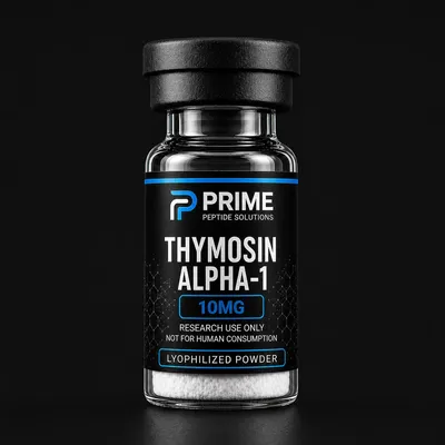
The Thymosin Alpha-1 in our catalog is supplied as a lyophilized powder in a sealed vial; the product page gives storage at 2-8 °C, or -20 °C for long-term storage. General peptide-handling guidance adds that for longer storage peptides are best kept as the lyophilizate in a tightly closed container below -15 °C, with lower temperatures preferred. Keep vials cold, dry and dark. Peptides tend to be hygroscopic, so a cold vial should reach room temperature in a desiccator before opening, and should be resealed tightly after weighing, because absorbed moisture lowers peptide content and may reduce stability.
Peptides containing Asn, Gln, Met, Cys or Trp have limited shelf lives; thymosin alpha-1 contains one Asn (its C-terminal residue) and none of the others. See our guide to storing research peptides and the lyophilization explainer.
Reconstitution
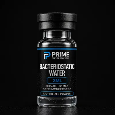
For solution work, the powder is reconstituted with bacteriostatic water (0.9% benzyl alcohol in sterile water), sold separately. The reconstituted solution is kept refrigerated at 2-8 °C, and general guidance advises against storing peptides in solution long-term. Final concentration equals the peptide mass in the vial divided by the volume of diluent added.
Verifying identity and purity
In general, identity and purity of a research peptide are documented on a batch Certificate of Analysis: HPLC separates the peptide from related impurities to give a purity figure, and mass spectrometry checks that the observed mass matches the expected value, about 3108 Da for thymosin alpha-1. Our guide to reading a peptide COA explains each field.
Frequently Asked Research Questions
What is thymosin alpha-1?
A 28-residue peptide with an acetylated N-terminus. It is the N-terminal segment (residues 2-29) of prothymosin alpha, and its sequence was first determined in 1977 from calf-thymus thymosin fraction 5.
Is thymosin alpha-1 the same as thymosin beta-4 or TB-500?
No. Thymosin alpha-1 comes from prothymosin alpha (gene PTMA), while thymosin beta-4 is encoded by TMSB4X, belongs to a different family and is annotated as actin-binding. They share only a historical name.
What receptor does thymosin alpha-1 act on?
A precise receptor has not been identified. Cell and animal studies report TLR9 and TLR2 signaling, with MyD88 as a key adaptor (TLR2 binding was measured directly by surface plasmon resonance, at micromolar affinity), and reviews also list TLR3, TLR4 and TLR7.
What is the research-grade thymosin alpha-1 sold here intended for?
The research-grade thymosin alpha-1 sold by Prime Peptide Solutions is not approved for human use. It is supplied only for in-vitro laboratory research and is not for human or animal consumption. It is not a medicine and should not be regarded as equivalent to any approved medicine.
Conclusion
Thymosin alpha-1 is a 28-residue, N-acetylated, highly acidic peptide cut from the N-terminus of prothymosin alpha, unstructured in water and helical in negatively charged model membranes (micelles and phospholipid vesicles). No precise receptor has been identified; work in cell and animal models centers on TLR/MyD88 signaling in dendritic cells, with downstream p38 MAPK/NF-kB, IRF7 and IDO pathways. It is unrelated to thymosin beta-4 despite the name. For laboratories, the practical points are cold, dry storage, refrigeration after reconstitution and identity checks against a mass of about 3108 Da.
Disclaimer: This article is provided for educational and research purposes only. It summarizes publicly available scientific literature and does not constitute medical advice. Thymosin alpha-1 and all peptide compounds sold by Prime Peptide Solutions are intended strictly for laboratory research, are not approved for human use, and are not for human or animal consumption. Researchers are responsible for compliance with all applicable regulations in their jurisdiction.
Shop these products
- Ships the same business day
- Free US shipping on $199+
- 14-day money-back guarantee
References & Further Reading
- PubMed: Current peer-reviewed thymosin alpha-1 literature
- PubMed: Thymosin alpha-1 and toll-like receptor signaling
- PubMed: Thymosin alpha-1 and dendritic cells
- PubMed: Prothymosin alpha research
- PubMed: Thymosin alpha-1 structure by NMR
- Related: Peptide Reconstitution Guide: Ratios and Storage
Research-Grade Thymosin Alpha-1
In our catalog, research-grade thymosin alpha-1 is listed as Thymosin Alpha-1, supplied as a lyophilized powder in one size, a 10mg vial, with bacteriostatic water sold separately. Batch Certificates of Analysis are published on the COA page as they become available; check it for a report matching the product and batch.
Sold strictly for in-vitro laboratory research. Not for human or animal consumption.