

What Should a Reconstituted Peptide Solution Look Like?
A lyophilized peptide that has fully dissolved should give a clear solution: see-through, with no haze, flakes, strands, film, gel or sediment. Clear means transparent, not colorless; GHK-Cu, for example, is blue because of its copper(II) complex. A milky, foamy or thick look can be temporary, such as trapped air, or it can be real aggregation. This guide covers the common causes, the limits of judging by eye, and when to discard a vial.
For inspection, a 2026 laboratory study wiped each vial with a dust-free tissue, swirled it for 5 seconds and inspected it against a black and a white background. Our bench routine borrows the idea (our advice, not the paper's protocol): wipe, swirl gently, let the vial rest so bubbles clear, then look against a dark and a light background.
This page covers appearance only. The full procedure is in our general reconstitution guide, and storage time is covered in the peptide stability guide.
This guide is a laboratory handling reference. Research-grade compounds from Prime Peptide Solutions are sold strictly for in-vitro laboratory research, are not approved for human use, and are not for human or animal consumption.
Foam and Microbubbles vs True Cloudiness
Why shaking matters
Shaking whips air into the liquid. A 2017 review of peptide aggregation describes air-water interfaces as hydrophobic, notes that amyloid-beta(1-40) and insulin both aggregate at them, and says agitation "is routinely used in studies of peptide and protein aggregation". In a 2008 study, shaking or stirring a monoclonal antibody solution produced aggregates; stirring was the harsher stress there, and vial headspace strongly influenced stability under shaking.
So add the diluent slowly down the vial wall, then swirl or roll gently. A few bubbles from gentle mixing settle and are not a discard reason. Heavy foam from shaking is avoidable mechanical stress.
Foam and a slowly dissolving cake
Foam can also form as a cake dissolves. In a 2021 study of high-concentration lyophilized protein cakes, some cakes floated "amidst foam or bubbles generated during reconstitution" and dissolved by gradual surface erosion, more slowly when viscosity near the surface was higher. The study reports this as an observation during reconstitution; it does not describe it as a sign of degradation. A floating cake needs time, not force.
Telling bubbles from haze
In our experience, microbubbles cling to the glass or drift upward and thin out on standing, while true turbidity is an even haze or opalescence through the liquid that does not clear on standing. The eye has limits: a flow-imaging paper lists air bubbles as particle look-alikes and notes that some sub-visible protein particles "can be highly transparent, fragile, and unstable". A clear-looking solution can still hold aggregates too small to see.
Why Solutions Turn Cloudy: Temperature, Concentration and pH
Cold diluent and slow dissolution
Water is more viscous when cold: NIST data give 1.5673 mPa·s at 4 °C against 1.0016 mPa·s at 20 °C, about 1.6 times as high. Since higher viscosity near a dissolving cake has been linked to slower reconstitution, cold diluent plausibly dissolves a cake more slowly, leaving a hazy, part-dissolved look for longer. A 2024 antibody study also named diluent temperature and swirling frequency as significant factors, but that is not a reason to warm vials or diluent. Let a cold vial reach room temperature, swirl gently and wait.
Cold-linked haze can also be physical and reversible. In a 2014 paper, one monoclonal antibody formed an opaque white gel above 30 mg/mL and below 6 °C that reversed with temperature; that is an antibody, not a peptide. Gas solubility also falls as temperature rises, so a warming solution can release fine bubbles onto the glass. Our rule: haze that does not clear after the vial rests and reaches room temperature is a discard signal.
High concentration
The 2017 review calls concentration "one of the most important factors influencing the physical stability of peptides": aggregation half-time and lag time generally shorten as it rises, and instability can "become a serious problem at high concentrations".
Nominal concentration is vial mass (mg) divided by diluent volume (mL), ignoring the small volume the dissolved solid adds. Halving the diluent volume doubles the concentration of the stock. Less diluent means a more concentrated, more aggregation-prone stock, and no solubility limits have been verified for these compounds.
pH, net charge and the isoelectric point
Net charge depends on pH, and charge repulsion must be overcome before molecules self-associate. In the review's words, "the lower the net charge the higher the propensity to aggregate", and near the isoelectric point (pI), where net charge is essentially zero, "monodisperse and quasi-amorphous aggregates" have been observed. A 2001 study of a small protein found that the pH of minimum solubility tracks the pI, and that calculated pI values can be off by more than one pH unit. So a pH near a peptide's pI favors haze, though a calculated pI is only an estimate. The review also notes that some self-assembling peptides form hydrogels, one possible explanation for a gel-like solution.
What about the preservative?
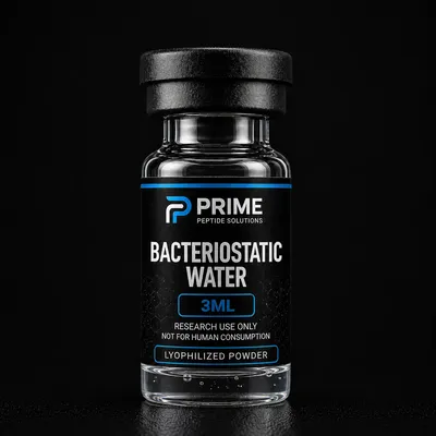
Our bacteriostatic water is 0.9% benzyl alcohol in sterile water. Preservative effects are molecule-specific: in a 2022 NMR study, benzyl alcohol did not aggregate an acylated 31-residue peptide, while 1% m-cresol produced insoluble aggregates after 24 hours at room temperature. Freezing adds a separate risk. In a 2026 study, benzyl alcohol worsened freeze-thaw aggregation of a model monoclonal antibody, and the authors recommended holding reconstituted solutions at 2-8 °C rather than freezing them.
Color Changes and Compound-Specific Signs
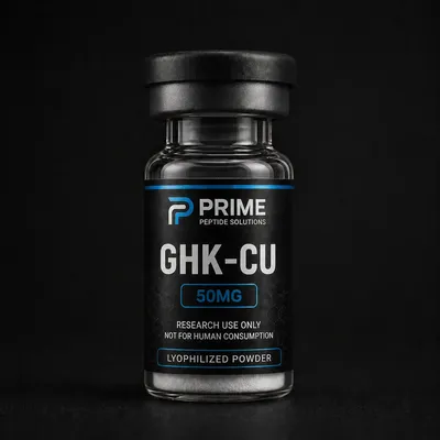
GHK-Cu: when the blue fades
GHK-Cu is blue because of copper(II), which absorbs orange-red light; Cu(I), with a full d-shell, has no such color. A 2021 study measured the Cu(II)-GHK band at 606 nm and saw it drop at once when reduced glutathione reduced the copper, then recover slowly as it re-oxidized. A 2026 review adds that pH changes how copper is bound, that phosphate and citrate buffers, chelators and proteins can pull copper away from GHK, and that reducing agents shift the Cu(II)/Cu(I) balance.
A paler solution after more diluent is simply more dilute. Fading in the same solution over time, or after mixing, points to changed copper chemistry, not necessarily peptide breakdown. Color is qualitative only: blue does not prove purity, and pale is not proof of a problem.
NAD+: any color change is a discard signal
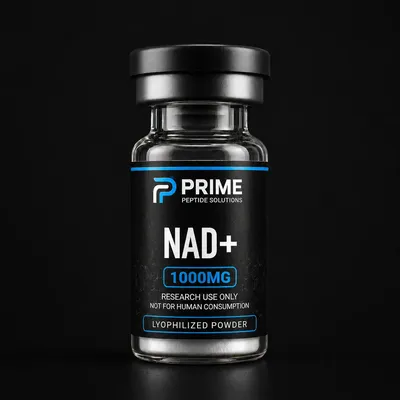
NAD+ is a dinucleotide coenzyme, not a peptide (C21H27N7O14P2, molecular weight 663.4 g/mol), and a highly hygroscopic white solid. The oxidized form undergoes base-catalyzed degradation at high pH, and the buffer also matters: in a 43-day study with each buffer at pH 8.5, a 2 mM solution degraded nearly entirely in HEPES, while in Tris its 260 nm absorbance fell only 4%. That was tracked by UV absorbance, not by eye, and none of the sources reviewed here describes a visible degradation color. Our rule: any color change, haze or particles in an NAD+ solution means discard.
IGF-1 LR3: charge, pI and solvent choice
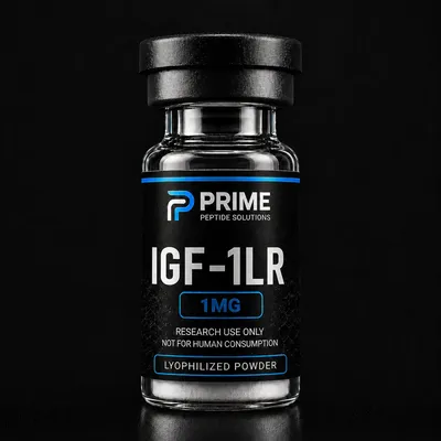
IGF-1 LR3 is an 83-residue analog of human IGF-I, with Glu-3 replaced by Arg and a 13-residue N-terminal extension; in cell models these changes alter its interaction with IGF-binding proteins, while IGF-I itself signals through IGF1R. Sequence-based calculation gives a theoretical pI of about 8.6 (native IGF-I: 7.76). That estimate may be off by a pH unit or more, but it suggests little net charge in the mildly basic range and a clear positive charge in dilute acid.
In published work, the stock for the cell-culture work in a 2025 study was made in 100 mM acetic acid following a supplier's protocol, while a 2026 dataset used water as the manufacturer-recommended buffer. Dilute acetic acid is a solvent used in published protocols, not the only option. The store does not sell acetic acid or any other acidic diluent; bacteriostatic water is the only diluent we carry.
When to Discard a Vial
Our handling rules, the same for every compound:
- Persistent haze: cloudiness or opalescence that does not clear after the vial rests and reaches room temperature.
- Visible particles: flakes, fibers, strands, specks, sediment, or a film on the glass or surface.
- Gel or thickening: a viscous, stringy or gel-like solution.
- Color change: any change in the same solution after it was freshly prepared, including a fading of GHK-Cu blue over time. A GHK-Cu solution that is paler only because more diluent was used is not a color change.
- Accidental freezing: a solution made with bacteriostatic water that froze. Replacing it is the conservative choice.
Do not try to rescue a cloudy vial by filtering, adding diluent, adding acid or base, or reheating. None of this is supported, and filtering removes material and changes the concentration.
A clear solution is not proof of an intact peptide either, since oxidation and hydrolysis are invisible. Identity and purity are established analytically, by the methods in our HPLC and mass spec explainer, not by eye.
Frequently Asked Research Questions
Can cloudiness right after mixing be temporary?
Yes. Trapped air and a cake that is still dissolving can look milky. Let the vial rest at room temperature and swirl gently. If haze, particles or gel remain, discard it.
Does cloudiness mean the material is impure or misidentified?
Not necessarily. Bubbles, reversible self-association, aggregation, high concentration and contamination can look alike, and the eye cannot reliably tell them apart. Apply the discard rule; identity and purity are established by HPLC and mass spectrometry, not by eye.
Is acetic acid required for IGF-1 LR3?
No single solvent is required. Dilute acetic acid appears in published protocols, and at least one dataset used water. The store does not sell acetic acid, and solvent choice belongs to the study protocol.
Conclusion
The causes covered here fall into two groups: a temporary look from bubbles, cold diluent or a still-dissolving cake, and real aggregation driven by agitation, high concentration or a pH near the pI. Contamination can look the same, and the eye cannot reliably tell these apart. Gentle swirling, patience and a room-temperature rest separate the two as well as the eye can. Haze, particles, gel or a color change that persists means discard, and a clear solution still relies on HPLC and mass spectrometry for identity and purity.
Disclaimer: This guide is for educational and laboratory reference only and is not medical advice. All compounds sold by Prime Peptide Solutions are intended strictly for in-vitro laboratory research, are not approved for human use, and are not for human or animal consumption. Researchers are responsible for complying with the regulations that apply in their jurisdiction.
Shop these products
- Ships the same business day
- Free US shipping on $199+
- 14-day money-back guarantee
References & Further Reading
- PubMed: Peptide aggregation and the isoelectric point
- PubMed: Agitation-induced protein aggregation
- PubMed: Cu(II)-GHK and thiol reduction
- PubMed: NAD+ stability in buffer solutions
- PubMed: Arg3 IGF-I analog literature
Research-Grade Peptides and COA Documentation
In our catalog, the compounds discussed here are listed as GHK-Cu (50mg and 100mg), IGF-1 LR3 (1mg) and the coenzyme NAD+ (1000mg), with bacteriostatic water sold separately in 3 mL and 10 mL vials. A batch COA for GHK-Cu is published on our COA page. For IGF-1 LR3 and NAD+, batch Certificates of Analysis are published on the COA page as they become available; check it for a report matching the product and batch.
Sold strictly for in-vitro laboratory research. Not for human or animal consumption.